



Eye Test at Vision in the Village
Comprehensive Eye Care in Southfields Village
Advanced Diagnostic Technology
What to Expect During Your Visit
Protecting Your Long-Term Eye Health
Book Your Eye Test Today
Frequently Asked Questions
What is the difference between OCT and Optomap scanning?
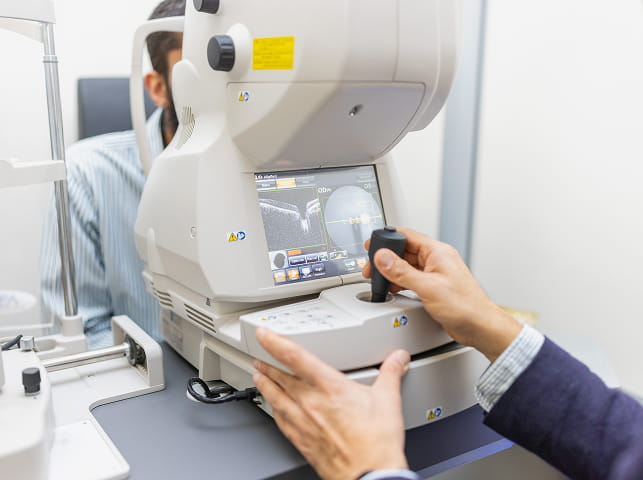
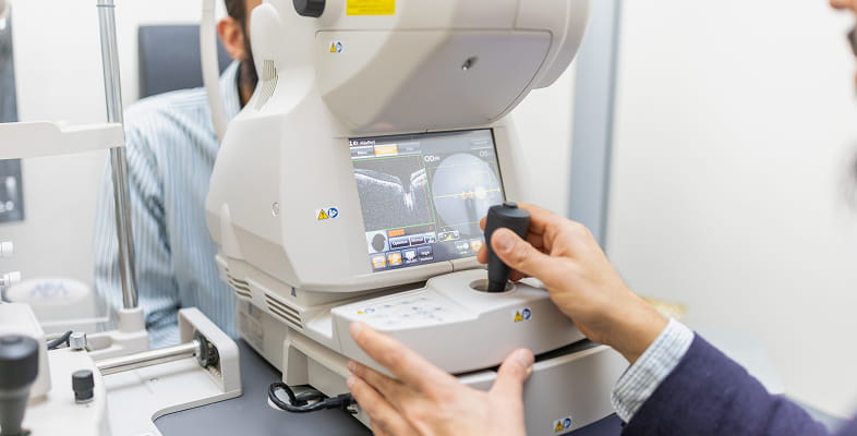
OCT (Optical Coherence Tomography) and Optomap scanning at Vision in the Village Opticians are complementary diagnostic technologies that examine different aspects of your eye health to provide comprehensive assessment. Our DRI OCT Triton captures ultra-high resolution cross-sectional images of your retina, similar to how an MRI scans body tissues, revealing individual cellular layers with exceptional clarity and measuring retinal thickness down to microscopic levels.
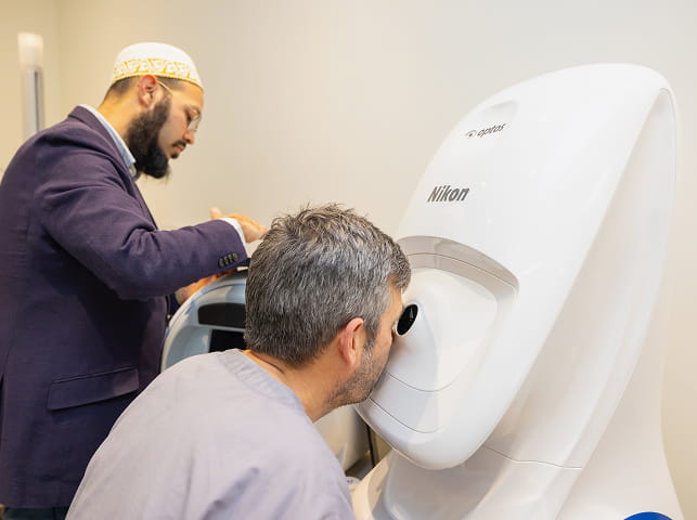
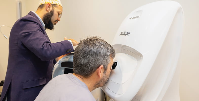
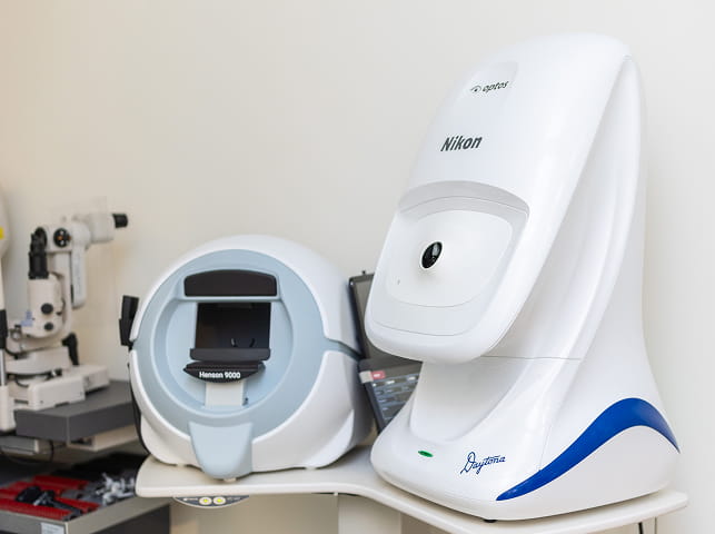
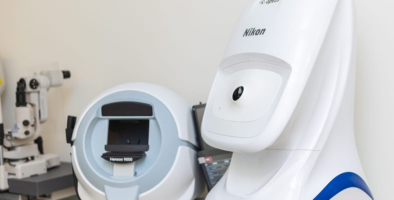
This swept-source OCT technology penetrates deeper into eye structures than conventional OCT systems, providing superior visualisation of the choroid (the vascular layer beneath the retina), the macula, and the optic nerve head, making it essential for detecting and monitoring glaucoma, age-related macular degeneration, diabetic macular oedema, and other conditions affecting retinal structure. In contrast, our Optomap Daytona provides ultra-widefield surface imaging, capturing over 80% of your retina in a single panoramic photograph compared to just 15% with traditional retinal photography.
This expansive view reveals peripheral retinal areas where serious conditions such as retinal tears, holes, lattice degeneration, and even ocular melanomas often develop unnoticed, as these areas lie beyond the reach of standard examination techniques. While OCT examines depth and layer structure in the central retina, Optomap examines breadth and surface features across the entire retinal landscape.
Together at Vision in the Village, these technologies work synergistically, OCT provides detailed structural analysis of the macula and optic disc, whilst Optomap ensures no peripheral pathology goes undetected. Both procedures are completely painless, non-invasive, take just seconds to complete, and usually require no dilating eye drops.
What can Optomap ultra-widefield imaging detect that standard photography cannot?
Optomap Daytona ultra-widefield imaging captures over 80% of your retina in a single panoramic image, providing crucial visibility of peripheral retinal areas that remain completely unseen with traditional retinal photography, which typically images only the central 15-30% of the retina.
This expanded view is clinically significant because many serious sight-threatening conditions develop in the retinal periphery, often progressing silently without symptoms until they affect central vision. Peripheral retinal tears and holes, which can lead to retinal detachment if left undetected, frequently occur in the far periphery beyond the reach of standard examination and photography, but are clearly visible on Optomap imaging, enabling timely referral for preventative laser treatment. Lattice degeneration areas of retinal thinning that predispose to tears and detachment is predominantly a peripheral condition that Optomap reveals comprehensively, allowing our optometrists to identify at-risk patients who require regular monitoring or prophylactic treatment.
Ocular melanomas and other retinal tumours often develop in the peripheral retina where they remain asymptomatic until reaching advanced stages; ultra-widefield imaging enables early detection when treatment outcomes are significantly better. Peripheral retinal haemorrhages and vascular abnormalities associated with diabetic retinopathy, retinal vein occlusions, and hypertensive retinopathy are frequently more extensive than central examination suggests, and Optomap imaging reveals the true extent of disease, informing more accurate grading and management decisions. The technology is particularly valuable for high myopes (very short-sighted individuals) who face increased risk of peripheral retinal pathology, and for patients with family history of retinal detachment.
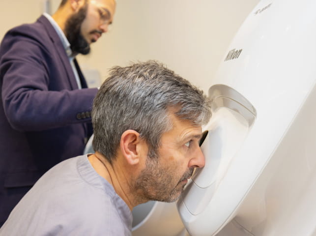
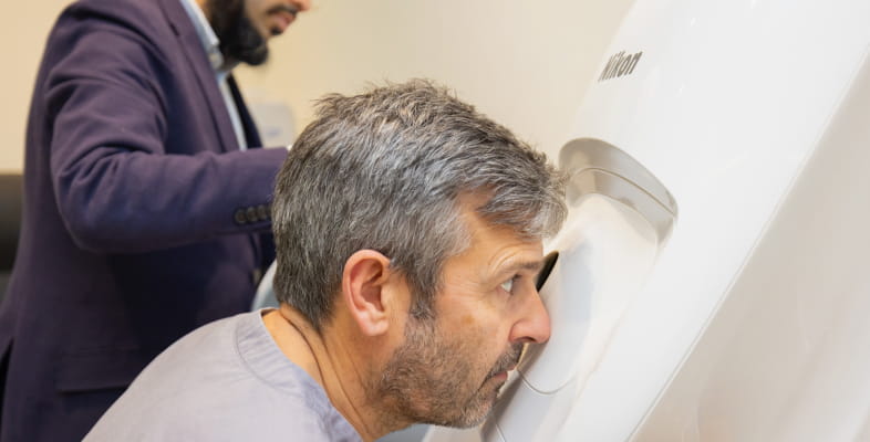
The Optomap scanning process at Vision in the Village is quick and comfortable, usually requiring no dilating drops, and produces detailed colour images that our optometrists can show you during your consultation, helping you understand your eye health clearly.
Do I need both OCT and Optomap scanning or just one?
We recommend both OCT and Optomap scanning as part of comprehensive eye examinations because these technologies provide complementary information that together creates a complete picture of your eye health that neither technology can achieve alone.
OCT scanning with our Triton system examines retinal structure in depth, revealing cross-sectional detail of cellular layers, measuring retinal thickness, and detecting subtle structural changes in the macula and optic nerve that indicate early glaucoma, macular degeneration, or diabetic macular oedema, conditions affecting the central retina that require detailed structural analysis.
Optomap ultra-widefield imaging examines retinal breadth, capturing surface features across 80% of the retina including peripheral areas where tears, holes, and other conditions develop beyond OCT’s scanning area and standard examination reach. Consider that a patient might have perfectly healthy central retinal structure on OCT but harbour a peripheral retinal tear visible only on Optomap, or conversely might show normal peripheral retina on Optomap whilst OCT reveals early glaucomatous nerve fibre layer thinning.
The clinical value of combining both technologies is well-established in optometric and ophthalmological practice, together they provide diagnostic confidence that reduces unnecessary hospital referrals whilst ensuring serious conditions never go undetected. For patients with diabetes, both technologies are essential: OCT detects and quantifies macular oedema requiring treatment, whilst Optomap reveals peripheral ischaemia and neovascularisation indicating disease progression.
During your consultation, our optometrists will discuss your individual risk factors and explain which imaging protocols are most appropriate for your circumstances, though most patients choose comprehensive screening with both technologies for complete peace of mind and clinical thoroughness.
How much does OCT and Optomap scanning cost at Vision in the Village?
Advanced diagnostic imaging at Vision in the Village Opticians is available as an enhancement to comprehensive eye examinations, with pricing structured to reflect the significant investment in hospital-grade technology and expert clinical interpretation.
OCT scanning with our DRI OCT Triton system is typically priced at £25-£35 when added to a comprehensive eye examination, providing detailed cross-sectional imaging of your retinal layers, macula, and optic nerve structures. Optomap Daytona ultra-widefield retinal imaging is generally priced at £25-£35, capturing panoramic images of over 80% of your retina including crucial peripheral areas. Many patients at our Southfields practice choose our combined advanced imaging package, which includes both OCT and Optomap scanning alongside a comprehensive eye examination conducted by our optometrists, with total appointment costs typically ranging from £85-£120 depending on the complexity of assessment required.
Your detailed scans are stored securely in your clinical records, creating valuable baseline measurements for future comparison and enabling our optometrists to detect even subtle changes during subsequent examinations. Some private health insurance policies cover advanced diagnostic imaging, so we recommend checking your policy details or contacting your insurance provider to determine coverage.
For accurate current pricing and to discuss which imaging services are most appropriate for your individual eye health needs and risk factors, please contact our friendly team at Vision in the Village directly we’re happy to provide detailed cost information and explain the clinical benefits of each technology based on your age, medical history, family history, and specific concerns.